Screening for Femoroacetabular Impingement Syndrome: What Are Our Options?

Femoroacetabular impingement syndrome (FAIS) was described as early as 1936. As this condition has become more recognized among the medical community, the number of patients identified with FAIS has significantly increased.
With this increased recognition, there has also been a marked rise in hip arthroscopy surgeries being performed. Literature has demonstrated an 18-fold increase from 1999 to 20091 and a 25-fold increase between 2006 and 2013.2
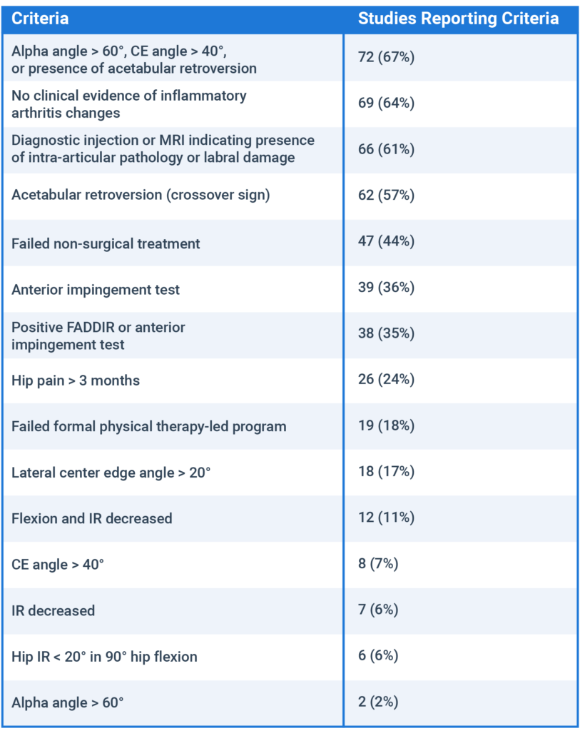
With regards to indication for surgery, Peters and colleagues performed a scoping review to identify what factors surgeons use to make this decision.3 This study found the following criteria used by surgeons for surgical intervention in the literature:

As you can see, the vast majority of the information used to determine surgery in this patient population is related to radiological findings and the extent of morphological changes. Most surprisingly, failure of conservative management and especially failure of formal physical therapy was not included in the vast majority of published studies.
This means that we might be getting ahead of ourselves.
What Is FAIS?
The femoroacetabular joint refers to the articulation between the proximal femur and the acetabulum of the pelvis. In FAIS, altered boney morphology of the femoral neck (cam morphology) or the acetabular rim (pincer morphology) leads to premature contact of the two osseous structures.
Based on the orientation of the joint, this premature contact typically occurs during hip flexion and/or internal rotation. This abnormal contact has also been blamed for additional pathological conditions such as acetabular labral tears, chondral lesions, and osteoarthritis.
Altered Morphology Does Not Always Matter
As with most morphological abnormalities, these factors do not always lead to pain and are fairly common in the general and athletic populations.
A systematic review conducted by Frank and colleagues of 2,114 asymptomatic hips found a very high prevalence of altered morphology. They found that 67 percent of subjects had radiologically confirmed pincer morphology, whereas 37 to 55 percent of athletes and 23 percent of the general population demonstrated cam morphology.4
To further evaluate the association of morphology in symptomatic patients, athletes, and asymptomatic individuals, Mascarenhas and colleagues performed a systematic review of 60 studies. This study found that cam morphology was significantly more common in athletes versus asymptomatic subjects but not compared to symptomatic patients, and significantly more common in symptomatic versus asymptomatic cases. No significant differences were found between pincer morphology prevalence when comparing athletes to symptomatic patients. However, mixed-type FAI was significantly more common in athletes versus asymptomatic subjects and in asymptomatic versus symptomatic subjects.5
Additionally, when looking at those pathologies that are said to be caused by altered morphology, the prevalence is also very high among asymptomatic individuals. The presence of acetabular labral tears and chondral lesions were found in asymptomatic individuals with a prevalence of 44 to 69 percent and 20 to 24 percent, respectively.6, 7
In fact, the level of morphological abnormality often does not coincide with severity of symptoms. A study of 500 adults found no association between radiographic signs of FAIS or a positive flexion adduction internal rotation (FADDIR) test with degree of hip pain.8 Jacobs and colleagues investigated the relationship of preoperative symptom severity and magnitude of boney morphology. This study of 64 patients prior to arthroscopic hip surgery found no correlation between symptom severity and degree of acetabular labral tear or femoroacetabular boney morphology. There was, however, a significant influence of depressive symptoms (as determined by the mental component score) and severity of hip-related symptoms,9 which gives further credence to the link between psychosocial factors and symptom severity regardless of morphological or pathological changes.
When Does Altered Morphology Matter?
Previous diagnostic criteria for femoroacetabular impingement relied heavily upon the level of morphological changes. Ganz, et al., and Sankar, et al., determined that the diagnosis of FAIS was appropriate if:
- There was abnormal morphology of the femur and/or acetabulum.
- There was abnormal contact between these two structures.
- The patient participated in activities with supraphysiologic motion, resulting in such abnormal contact and collision.
- The repetitive motion resulted in the continuous insult.
- Soft tissue damage was present.10, 11
Once again, these criteria are not sufficient to accurately diagnose a patient with FAIS because there is no weight put on clinical signs or symptoms.
More recently, Griffin and colleagues attempted to better define appropriate terminology, diagnosis, treatment, and prognosis for FAIS. At this consensus meeting, they agreed that accurate diagnosis depended upon clinical signs, symptoms, and diagnostic criteria. They therefore defined FAIS as “a motion-related clinical disorder of the hip with a triad of symptoms, clinical signs, and imaging findings. It represents symptomatic premature contact between the proximal femur and the acetabulum.”12
The key differentiating factors between this and previous descriptions are the additional criteria of ‘symptomatic’ and the emphasis on symptoms and clinical signs in addition to diagnostic criteria. This definition was met with a 9.8/10 agreement and allows for the entire patient presentation to be taken into consideration, not just the underlying morphological changes.
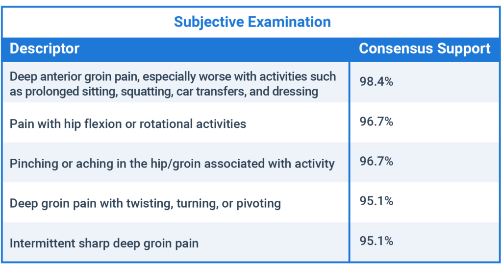
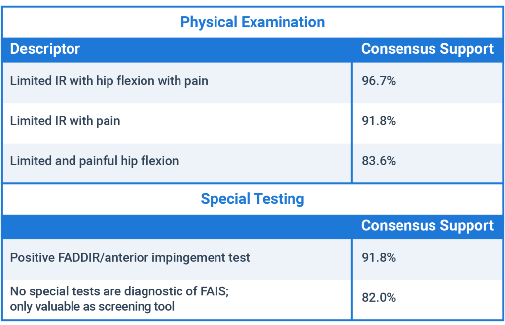
To expand upon this agreement, Reiman and colleagues performed an international and multi-disciplinary Delphi survey to identify pertinent aspects of the subjective history, clinical examination, and radiological examination.13 This survey found agreement on the following aspects in patients presenting with FAIS:

Subjective self-report should be the cornerstone of the examination of any injury, and FAIS is no exception. Patients often present with reports of deep anterior groin pain exacerbated by activities involving deep flexion, rotation, and squatting.
The subjective attributes agreed upon for individuals presenting with FAIS closely coincide with a diagnostic study performed by Clohisy and colleagues who looked at 51 patients with confirmed, symptomatic FAIS. This study showed that 88 percent of patients had pain localized to the groin region. Aggravating factors included general activity-related (71 percent), running (69 percent), sitting (65 percent), and pivoting (63 percent).14

A systematic review of 16 studies related to physical impairments in individuals with FAIS demonstrates similar findings as the Delphi survey. This study agreed that the available literature currently demonstrates that individuals with FAIS have decreased hip ROM into impingement (flexion/internal rotation in 90° flexion), which is often limited by pain.15
Consolidating Contradictory Criteria
When looking at the included criteria for special testing in FAIS, the two agreed-upon findings seem contradictory. On one hand, a positive FADDIR test is beneficial; on the other hand, it is also noted that no special tests are diagnostic for FAIS.
According to the literature in reference to special testing for FAIS, there has been no test that can be seen as confirmatory of the diagnosis due to very low positive likelihood ratios and specificity values.16 That being said, the use of the FADDIR test does offer benefit due to the very high sensitivity and low negative likelihood ratios reported in the literature (Sn = 0.94-0.99, -LR = 0.14-0.45);17 however, its capacity as a screening method has recently come into question.
A cross-sectional study of 74 ice hockey players (average age of 16 years old) contradicted the current literature with regards to the FADDIR test’s screening capacity.18 This unique study questions the FADDIR test’s capacity to screen for pure cam, pincer, or combined morphology (Sn = 0.41, -LR = 1.24) and pure cam or combined morphology (Sn = 0.60, -LR = 0.78).
As we continue to evaluate the capacity to screen for FAIS, there will be more consensus, but as of now the FADDIR can be used as a screening tool with caution, taking into consideration the patient’s additional clinical signs and subjective complaints.
Understanding that FAIS is far more than morphological changes to the proximal femur or acetabulum will allow us as clinicians and researchers to move forward in the evaluation, treatment, and return to sport of this patient population. By evaluating conservative management in returning athletes to their prior level of function, this drastic spike in surgical procedures may start to stabilize.
Boney morphology is a well-known contributor to FAIS, but it only tells one portion of the story. We need to dig deeper in order to successfully manage this patient population.
- Palmer, A. J. R., Malak, T. T., Broomfield, J., Holton, J., Majkowski, L. Thomas, G. E. R., & Taylor, A., et al. (2016). Past and projected temporal trends in arthroscopic hip surgery in England between 2002 and 2013. BMJ Open Sport & Exercise Medicine, 2(1), e000082.
- Cvetanovich, G. L., Chalmers, P. N., Levy, D. M., Mather 3rd, R. C., Harris, J. D., Bush-Joseph, C. A., & Nho, S. J. (2016). Hip arthroscopy surgical volume trends and 30-day postoperative complications. Arthroscopy, 32(7), 1286–92.
- Peters, S., Laing, A., Emerson, C., Mutchler, K., Joyce, T., Thorborg, K., & Hömlich, P., et al. (2017). Surgical criteria for femoroacetabula impingement syndrome: a scoping review. British Journal of Sports Medicine, 0, 1–6.
- Frank, J. M., Harris, J. D., Erickson, B. J., Slikker 3rd, W., Bush-Joseph, C. A., Salata, M. J., & Nho, S. J. (2015). Prevalence of femoroacetabular impingement imaging findings in asymptomatic volunteers: a systematic review. Arthroscopy, 31(6), 1199–204.
- Mascarenhas, V. V., Rego, P., Dantas, P., Morais, F., McWilliams, J., Collado, D., & Marques, H., et al. (2016). Imaging prevalence of femoroacetabular impingement in symptomatic patients, athletes, and asymptomatic individuals: a systematic review. European Journal of Radiology, 85(1), 73–95.
- Tresch, F., Dietrich, T. J., Pfirrmann, C. W. A., & Sutter, R. (2016). Hip MRI: Prevalance of articular cartilage defects and labral tears in asymptomatic volunteers. A comparison with a matched population of patients with femoroacetabular impingement. Journal of Magnetic Resonance Imaging, 46(2), 440–451.
- Register, B. Pennock, A. T., Ho, C. P. Strickland, C. D., Lawand, A., & Philippon, M. J. (2012). Prevalence of abnormal hip findings in asymptomatic participants: a prospective, blinded study. The American Journal of Sports Medicine, 40(12), 2720–2724.
- Kopec, J. A., Hong, Q., Wong, H., Zhang, C. J., Ratzlaff, C., Cibere, J., & Li, L. C., et al. (2020). Prevalence of femoroacetabular impingement syndrome among young and middle-aged white adults. Journal of Rheumatology, 47(9), 1440–1445.
- Jacobs, C. A., Burnham, J. M., Jochimsen, K. N., Molina 4th, D., Hamilton, D. A., & Duncan, S. T. (2017). Preoperative symptoms in femoroacetabular impingement patients are more related to mental health scores than the severity of labral tear or magnitude of bony deformity. Journal of Arthroplasty, 32(12), 3603–3606.
- Ganz, R., Parvizi, J., Beck, M., Leunig, M., Nötzli, H., & Siebenrock, K. A. (2003). Femoroacetabular impingement: a cause for osteoarthritis of the hip. Clinical Orthopaedics and Related Research, 417, 112–120.
- Sankar, W. N., Nevitt, M., Parvizi, J., Felson, D. T., Agricola, R., & Leunig, M. (2013). Femoroacetabular impingement: defining the condition and its role in the pathophysiology of osteoarthritis. Journal of the American Academy of Orthopaedic Surgeons, 21(Suppl 1), S7–S15.
- Griffin, D. R., Dickenson, E. J., O’Donnell, J., Agricola, R., Awan, T., Beck, M., & Clohisy, J. C., et al. (2016). The Warwick Agreement on femoroacetabular impingement syndrome (FAI syndrome): an international consensus statement. British Journal of Sports Medicine, 50, 1169–1176.
- Reiman, M. P., Thorburg, K., Covington, K., Cook, C. E., & Hölmich, P. (2017). Important clinical descriptors to include in the examination and assessment of patients with femoroacetabular impingement syndrome: an international and multi-disciplinary Delphi survey. Knee Surgery, Sports, Traumatology, Arthroscopy: Official Journal of the ESSKA, 25(6), 1975–1986.
- Clohisy, J. C., Knaus, E. R., Hunt, D. M., Lesher, J. M., Harris-Hayes, M., & Prather, H. (2009). Clinical presentation of patients with symptomatic anterior hip impingement. Clinical Orthopaedics and Related Research, 467(3), 638–44.
- Diamond, L. E., Dobson, F. L., Bennell, K. L., Wrigley, T. V., Hodges, P. W., & Hinman, R. S. (2015). Physical impairments and activity limitations in people with femoroacetabular impingement: a systematic review. British Journal of Sports Medicine, 49(4), 230–42.
- Caliesch, R., Sattelmayer, M., Reichenbach, S., Swahlen, M., & Hilfiker, R. (2020). Diagnostic accuracy of clinical tests for cam or pincer morphology in individuals with suspected FAI syndrome: a systematic review. BMJ Open Sport & Exercise Medicine, 6, e000772.
- Reiman, M. P. & Thorborg, K. (2014). Clinical examination and physical assessment of hip joint-related pain in athletes. International Journal of Sports Physical Therapy, 9(6), 737–755.
- Casartelli, N. C., Brunner, R., Maffiuletti, N. A., Bizzini, M., Leunig, M., Pfiffmann, C. W., & Sutter, R. (2018). The FADIR test accuracy for screening cam and pincer morphology in youth ice hockey players. Journal of Science and Medicine in Sport, 21(2), 134–138.


