Lights and Road Maps of Neuroscience: Exploring Brain Mapping Therapy

Advancements in neuroscience, particularly in brain mapping therapy, have revolutionized our understanding of the human brain. This therapy leverages cutting-edge imaging technologies to create detailed maps of brain activity and connectivity, aiding in the diagnosis and treatment of neurological conditions. These advancements are crucial for developing targeted rehabilitation programs for patients with brain injuries.
Understanding Brain Mapping Therapy
Brain mapping therapy is an innovative approach that involves using advanced imaging technologies like functional magnetic resonance imaging (fMRI) and Diffusion Tensor Imaging (DTI) to create detailed maps of the brain. These maps help clinicians and researchers understand the complex networks within the brain, allowing for more precise diagnoses and targeted treatment plans. This therapy is particularly useful in treating conditions like stroke, traumatic brain injuries, and neurodegenerative diseases, where understanding the brain’s structure and function is crucial for recovery.
Illuminating Brain Activity with fMRI
fMRI has been a cornerstone of neuroscience research since its inception at the turn of the century. fMRI, which stands for functional magnetic resonance imaging, sparked a neuroscience explosion by showing what parts of the brain “light up” when humans actively perform specific cognitive tasks. The research revealed that all cognitive functions occur through distributed networks of regions firing simultaneously.
However, this provided limited value because many areas involved in one cognitive task were multitasking—participating in many other, sometimes very different, cognitive activities. For example, regions of the inferior frontal lobe were active in both language and mathematical computation.
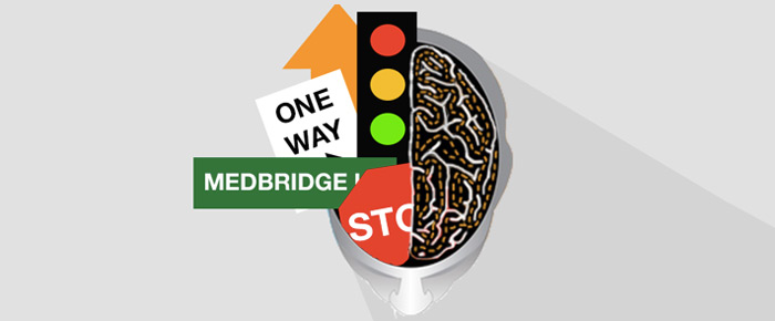
fMRI provided a geographical map of brain regions that were simultaneously active, similar to viewing activity in cities as you fly over them in a plane at night. You see lights but don’t know what the areas actually do or how the disparate regions are interconnected. This mapping of active regions during specific tasks laid the groundwork for brain mapping therapy by identifying the regions involved in various cognitive functions, which is crucial for targeted therapeutic interventions.
Visualizing the Brain with DTI
DTI emerged in the early 21st century, further advancing our understanding of brain connectivity. This technology enabled researchers to visualize the road map of the brain by clarifying fiber tracts that linked network regions together and by showing how maturation and experience are integral in building the human brain’s highway system. According to a study by Bagheri et al. (2023) on brain effective connectome, DTI tracks the movement of water molecules along white matter tracts, providing insights into the brain’s structural connectivity.
Yet, even with DTI, researchers were still unsure of the role individual regions played in cognitive processing. Researchers started measuring single neuron responses in specific brain regions — like Broca’s area and Wernicke’s area — to determine the exact type of stimulus those neurons are mapped to handle. This detailed mapping of neuron activity helped link specific brain regions to distinct cognitive functions, paving the way for targeted therapeutic interventions.
Recent Advances in Neuroscience Research
Recent research in neuroscience has focused on combining fMRI and DTI to develop more accurate brain maps. These maps help explain how damage to specific areas affects overall brain function. For instance, a study by Yen, Lin, and Chiang (2023) demonstrated how integrating fMRI and DTI data could help identify disruptions in brain networks caused by traumatic brain injuries. This research is designed to help therapists guide cognitive therapy for brain-injured adults by rebuilding the connectomes—the intricate web of neural connections that enable cognitive function.
Brain mapping therapy uses these insights to create individualized treatment plans. By understanding the specific damage in a patient’s brain and identifying the remaining healthy pathways, therapists can develop targeted rehabilitation programs that maximize recovery and improve the quality of life.
The Role of Brain Mapping Therapy in Rehabilitation
One of the primary applications of brain mapping therapy is in cognitive rehabilitation for stroke and traumatic brain injury patients. Using the detailed maps generated from fMRI and DTI, therapists can identify which areas of the brain are functioning normally and which are impaired. This allows them to tailor exercises and activities that stimulate the affected areas, promoting neural plasticity and recovery. For example, in stroke rehabilitation, brain mapping can help pinpoint areas that need targeted therapy to regain lost functions, such as speech or motor skills (Stinear et al., 2008).
In addition to cognitive rehabilitation, brain mapping therapy is showing promise in managing neurodegenerative diseases such as Alzheimer’s and Parkinson’s. By tracking the progression of these diseases and their impact on brain connectivity, therapists can create more effective intervention strategies that slow cognitive decline and improve patients’ quality of life.
Innovations and the Future of Brain Mapping Therapy
The future of brain mapping therapy looks promising, with ongoing research aimed at refining these techniques further. Scientists are exploring ways to integrate real-time data from fMRI and DTI into therapeutic protocols, providing immediate feedback to both therapists and patients. This integration could revolutionize cognitive rehabilitation by enabling more precise and individualized treatment plans, improving patient outcomes. A deeper understanding of cognitive functions and their neurological underpinnings can empower clinicians to apply these advancements more effectively in their practice.
To enhance your knowledge of cognitive functions and evidence-based interventions, explore the MedBridge course “Cognitive Interventions: Neuroscience Applications in Clinical Practice.” This course offers valuable insights into how neuroscience research can inform and improve clinical approaches to cognitive rehabilitation.
References
- Bagheri, A., Dehshiri, M., Bagheri, Y., Akhondi-Asl, A., & Nadjar Araabi, B. (2023). Brain effective connectome based on fMRI and DTI data: Bayesian causal learning and assessment. PloS one, 18(8), e0289406. https://doi.org/10.1371/journal.pone.0289406
- Yen, C., Lin, C. L., & Chiang, M. C. (2023). Exploring the Frontiers of Neuroimaging: A Review of Recent Advances in Understanding Brain Functioning and Disorders. Life (Basel, Switzerland), 13(7), 1472. https://doi.org/10.3390/life13071472
- Eliassen, J. C., Boespflug, E. L., Lamy, M., Allendorfer, J., Chu, W. J., & Szaflarski, J. P. (2008). Brain-mapping techniques for evaluating poststroke recovery and rehabilitation: a review. Topics in stroke rehabilitation, 15(5), 427–450. https://doi.org/10.1310/tsr1505-427

